Key Takeaway
Table of Contents
Normal Radiographic Anatomy for the INBDE
Understanding what normal radiographic anatomy is important in order to be able to identify when a radiographic finding is abnormal.
Tooth Anatomy

We can zoom in and see some additional structures:

Additional landmarks:

Some additional pulp features:

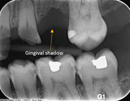
In some radiographs, the gingiva can be visible. This will appear as:
Additional supporting structures include cancellous/trabecular bone and cortical bone:

Maxillary Region
There are several other anatomic features that can be recognized in maxillary periapicals.
Intermaxillary suture
- Linear or slightly undulating radiolucent line between maxillary central incisors


Lateral fossa
- Diffuse radiolucent region apical and overlying maxillary lateral incisor


Canine fossa
- Diffuse radiolucent region distal to the maxillary canine

Anterior nasal spine
- diffuse , v-shaped radiopaque region apical to maxillary central incisors


Incisive foramen
- Oval radiolucency in region of maxillary central incisors


Nasopalatine canal
- Radiolucent channel between maxillary central incisors


Nasal septum
- Vertical radiopaque line at midline superior to central incisor


Nose
- Outline of soft tissue


Nasolabial fold
- Outline of soft tissue


Zygomatic arch
- Broad, radiopacity projected mostly superior and posterior to maxillary molars


Zygomatic process of maxilla
- U-shaped, radiopaque line in region of maxillary molars


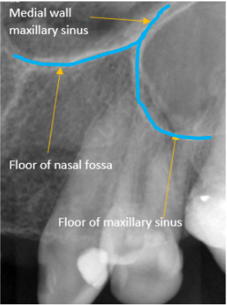
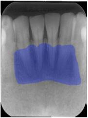
Floor of maxillary sinus
- Bottom portion of the pyramidal-shaped cavity above the roots of the maxillary premolars and molars


- The nasal cavity and sinuses can have many variations
- The nasal cavity and sinus can be normal, not visible, or large
- The paranasal sinuses can vary in size, shape, and internal septations
- The sinuses have many variants related to their outline, shape, and internal appearance
- The size of the maxillary sinus increases as an individual ages until skeletal maturity is gained
This characteristic “Y” shape is commonly tested!

Maxillary tuberosity
- Radiopaque, rounded prominence distal to maxillary molars


Hamulus
- Hook-like, radiopaque process projected posterior to maxillary tuberosity


Mandibular Region
Periapical radiographs in the mandibular posterior region can include anatomic features of the mandible.

Genial tubercles
- Poorly defined, irregular, radiopaque region apical to mandibular central incisors


Mental ridge
- Radiopaque, triangular shaped area below the mandibular anterior teeth


Mental foramen
- Round or oval radiolucency in apical region of mandibular premolars or first molars

- Variant of normal: two on one side or posteriorly positioned (this could be real or tube angulation)
Submandibular gland fossa
- Poorly defined, broad radiolucency apical to mandibular molars and premolars


Inferior Alveolar Canal
- Curvilinear, radiolucent channel in posterior mandible


- Variant of normal: anterior looping (shown below), multiple canals, canal diameter large or small

Mental fossa
- Poorly defined, broad, radiolucent region superimposed on mandibular incisor roots

Nutrient canals
- Radiolucent lines extending between tooth roots


Lingual Foramen
- Round, radiolucent point apical to mandibular central incisors

Sialolith
- Round or oval-shaped radiopacity located on the inside of the mandible
- Due to the calcification and formation of stones in the major salivary glands

Mandibular tori
- Broad radiopacity overlaying the roots of posterior mandibular teeth


Cervical burnout
- This term refers to the radiolucency found just below the CEJ on the root due to anatomical variation or a gap between the enamel and bone covering the root
- It can appear similarly to caries


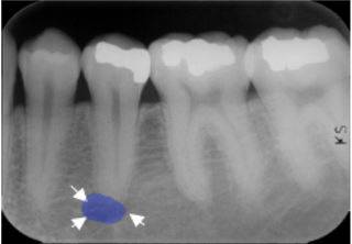
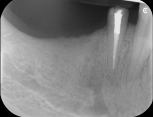
Sinus pneumatization
- The continuous physiological process that causes the paranasal sinuses to increase in volume
- Although it it seems like there is pathology between the roots of tooth 3 and 5, it is just the expanded sinus


Panoramic Features
There are several common anatomic features recognizable in the panoramic radiograph.


- Mandibular ramus- Broad radiopaque region in the mandible that connects the body to the condyle and coronoid process
- Mandibular angle- Junction of the posterior and inferior border of the mandibular ramus
- Condyle- Oval radiopaque process that extends posteriorly and superiorly from the mandibular ramus
- Coronoid process- Triangular radiopaque process that extends anterior and superiorly from the ramus
- Sigmoid notch- Concavity between the condyle and coronoid process
- Antegonial notch- Concavity on inferior border of the mandible
- Cervical spine- Irregular, radiopaque column behind the mandible and maxilla
- Hyoid- Radiopaque process inferior or even superimposed onto the mandible
- External auditory meatus- Radiolucent oval behind the mandibular condyle
- Styloid process- Pointed radiopacity behind the mandibular ramus
- Glenoid fossa- Thin radiopaque concavity where the condyle meets
- Articular eminence- Radiopaque convexity in front of the glenoid fossa
Soft tissue features can also superimpose onto the panoramic radiograph.


- Tongue- Creates an arch that superimposes over the mandibular teeth and portions of the maxillary teeth
- Soft palate- Creates an arch that superimposes above the tongue
- Epiglottis- Radiopaque structure seem behind the tongue
- Oropharynx- Creates a column of soft tissue that superimposes behind the mandible
- Nasopharynx- Creates a column of soft tissue that superimposes behind the mandible and is located above the oropharynx
Now that you know what normal looks like, make sure to check out INBDE Bootcamp's Radiographic Bone Lesions To Know!
Read This Next


%20(1)%20(1).jpg)
Save 100+ hours of your life studying with Bootcamp.com
Get everything you need in one place. Start studying today for free.